Characteristics and principle of the method
Magnetic resonance imaging (MRI) is one of the modern imaging methods. It is distinguished by its excellent contrast resolution of individual tissues, i.e. the ability to distinguish even tissues with very similar structures. In practice, this means not only, for example, excellent differentiation of the white and grey matter of the brain, but (more importantly) differentiation of normal tissue from tissue affected by a disease process. In this respect, magnetic resonance imaging has a privileged position among all imaging methods.
Translated with DeepL.com (free version)
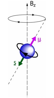
The principle of MRI is different from other imaging methods. It uses the specific physical properties of the nuclei of hydrogen atoms. Hydrogen nuclei, when exposed to a strong magnetic field, are the source of radiofrequency waves. These waves (among other things, very similar to the waves used to transmit radio signals in the VHF band) are picked up by a system of receiving coils (antennas).
It is important to mention that no harmful side effects of magnetic resonance imaging on the human body have been proven so far, it is possible from the 4th month of pregnancy!
The price of 1 examination in the Czech Republic is currently around 7 000 CZK.
Examination procedure
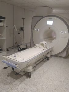
The examination begins with placing the patient on the examination table. The instruments are divided into closed and open. In the former, the patient is placed in a relatively confined space, which can be uncomfortable, especially for claustrophobic patients. The advantage, however, is the possibility of taking better quality images in a shorter time, as these devices have a stronger magnetic field.
During the examination, a contrast agent may be injected into a vein. Contrast agents used for MRI most often contain gadolinium compounds, rarely manganese or iron. The risk of an allergic reaction to these substances is extremely low. MRI does not use contrast agents containing iodine.
The duration of the MRI scan is in most cases between 20 and 50 minutes.
Preparation for examination
Preparation is practically not necessary. The patient does not need to be hungry before the intravenous administration of the contrast agent. Claustrophobic patients are advised to come to the examination with a companion (even a strong feeling of claustrophobia can be easily removed by applying a light sedative, but you must not drive or perform activities requiring concentration after the application).
If you are wearing any electronic or metallic implant or foreign body, it does not automatically mean that you cannot have an MRI scan. However, you must ALWAYS and BEFORE the examination report this fact to the operator of the MRI machine who will make an informed decision as to whether or not you can have the examination.
Use of the method (indications)
The use of MR is very wide, from classical imaging of the central nervous system to imaging of blood vessels (MR angiography), joints, organs of the chest (heart) and abdomen or other special techniques such as MR spectroscopy, functional MR of the brain, MR diffusion imaging, etc.
When the examination is not appropriate (contraindications)
MRI scans cannot be performed on people with certain types of electronic or metal implants or foreign bodies. In particular, patients with a pacemaker or implanted cardioverter defibrillator (ICD) cannot be examined because there is a risk of serious impairment of its function or serious or even life-threatening disturbance of the heart rhythm. Persons with metallic vascular clamps after cerebral artery aneurysm (aneurysm) surgery can be examined by MRI only under strictly specified conditions.

Where the examination is performed
Examinations are performed by specialized accredited radiological workplaces, our workplace is accredited by the Radiological Society of ČLS JEP, accredited by the SAK of the Czech Republic and ISO 9001:2001 quality certificate within the whole Hospital Jihlava.
You can make an appointment / inquire about an examination by calling 567 157 281


Leave a Reply
You must be logged in to post a comment.