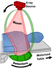
It is a device that works with X-rays in a similar way to a conventional X-ray machine, but in CT (computed tomography) the radiation source - the X-ray tube and the detection system - rotates around the patient's body, which is then irradiated gradually from different angles around the body. The detection system then evaluates the amount of radiation passing through the patient's body at different angles and from this data, individual scans - images of the body layers, usually in the axial plane - are then reconstructed using a powerful computer.
In a classical X-ray, the result is a summative image of the whole body in which the shadows of individual structures overlap; in a CT scan, we obtain thin layers of the examined area, without overlapping individual parts of the body, so it is possible to show individual organs, their structure and pathologies much more accurately.
Indications for examination
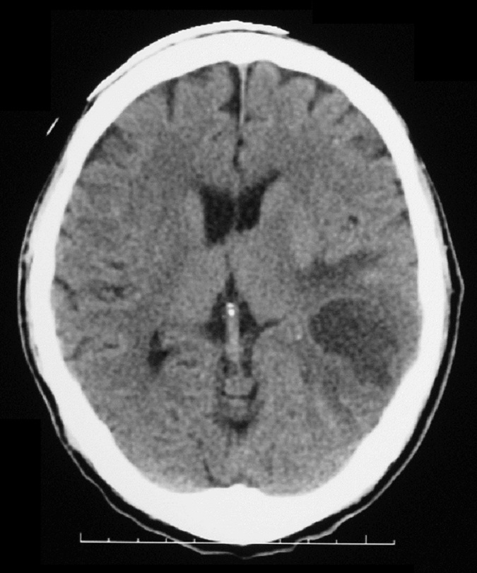
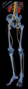
Due to the relatively high burden of X-rays, CT is indicated as a "second choice method" - a complementary examination that should help to clarify unclear findings of ultrasound or conventional X-rays, especially in the assessment of bones, lungs and intestines, where ultrasound is not applicable. For acute (urgent) indications, we use CT mainly for head-brain imaging in stroke and head trauma as it will show intracranial haemorrhage very well. Traumatic changes of the chest, abdomen, pelvis and bone fractures are also very reliable. Another very important acute indication for CT scanning is suspected aortic (heart) involvement, such as an aneurysm or dissection (tearing of the lining) of the aorta. In this case, CT angiography (CT examination of blood vessels) is performed, mainly to image the large arteries (aorta, renal arteries, pelvic arteries, carotid arteries) and also to image the cerebral arteries (circulus arteriosus Willis). In some cases, it can completely replace conventional angiographic examination.
Contraindications to the examination
Before the examination, it is necessary to make targeted inquiries and to exclude or confirm the presence of contraindications. Each patient is instructed by the attending physician about the risks of intravenous contrast agent administration and, if it is anticipated that it will be administered during the CT scan, the attending physician requests and countersigns the patient's written informed consent for its intravenous administration.
For relative contraindications, the risk-benefit ratio of the test must be considered.
Native examination
Gravity - only in serious danger to life.
Examination with a contrast agent
The examination must not be performed in patients allergic to iodine contrast agents.
In the cases listed below, a contrast CT scan may be performed only in emergency cases (vital indications - the scan must be performed to save the patient's life) and under emergency measures specific to the case (thorough and extended anti-allergic preparation, assistance of the anaesthesia team, provision of haemodialysis, necessary hospitalisation, etc.):
• pregnancy,
• hepatic and renal insufficiency, followed by dialysis,
• hyperthyroidism (increased thyroid function),
• pheochromocytoma (catecholamine-producing tumor),
• an uncooperative patient.
Risks of examination
The CT machine uses X-rays and therefore there is a certain radiation load on the patient, see section "Ionizing radiation".
Administration of a contrast agent (intravenously or orally) - may cause adverse effects: hot flushes, sweating, nausea, redness, itching, rash and, in very rare cases, a severe allergic reaction in the form of anaphylactic shock may occur, requiring medical attention and, exceptionally, hospitalisation of the patient.
You can make an appointment / inquire about an examination by calling 567 157 561
Instrumentation and sample images:
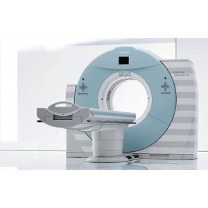
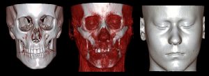
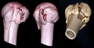
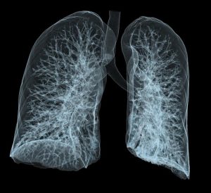
- CT Siemens SOMATOM DEFINITION AS+ 128CT The Siemens Somatom Definition AS+ allows simultaneous scanning of 128 layers per turn of the X-ray tube. The exceptional spatial resolution offers imaging of tissue structures as small as 0.24 mm, and the image scanning range of 200 cm combined with this resolution enables examination of the entire patient body in 10 seconds. This is particularly important when examining patients with severe trauma, unconscious or uncooperative patients. These exceptional parameters are particularly useful in traumatology, where it is important to make a diagnosis as quickly as possible, and in cardiology, where they allow examination of cardiac function and evaluation of coronary arteries, including analysis of calcifications in their walls.The speed and quality of the images are advantageous for qualitative vascular diagnosis and planning of interventional procedures on blood vessels. Fully automatic 3D volume imaging facilitates the evaluation of cerebral blood flow. Images from this device are generated in DICOM format, allowing immediate transmission to the PACS system and viewing of the submitted CT images at clinical sites.

Leave a Reply
You must be logged in to post a comment.